
Lung Disease Imaging with X-ray Interferometry
Interferometry detects the air-tissue interface in healthy alveoli
A Paradigm Shift in Diagnostic Lung Imaging
Current practice is an expensive CT scan
Interferometry offers more diagnostic information in less time and with less radiation exposure. A first-generation X-ray interferometer built at the Technical University of Munich has examined more than 500 COPD patients. Refined Imaging is developing a second-generation X-ray interferometer, with NIH STTR support, and planned clinical evaluations at the LSU Pennington Biomedical Research Center.
Overcoming the Limitations of Computed Tomography
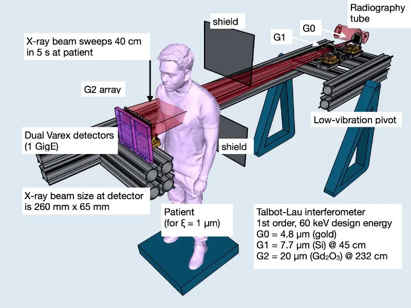
Second-generation X-ray interferometer will have walk-in patient access and seven second scan time.
In healthcare, the existing diagnostic standard for detecting lung diseases, cancer, and other maladies is computed tomography (CT) technology, which has three major drawbacks:
1) The image detail is not sufficient for effective early-stage diagnosis
2) Patients are exposed to substantial amounts of radiation and
3) CT is expensive, and accessibility is often limited
To solve these problems, Refined Imaging combines data analysis software and hardware (microfabricated X-ray optics) to eliminate the current drawbacks of CT scanning.
Understanding Grating-Based Interferometry
X-ray interferometry adds phase contrast and scattering images onto conventional absorption image.
Conventional X-ray imaging uses absorption to show soft tissue contrast, which is insufficient for important organs such as the lung, liver, uterus, and prostate.
With X-ray interferometry, diagnosticians gain simultaneous access to three complementary and correlated images in a single scan:
- A scattering image showing X-ray interaction with sub-micron tissue structures,
- A differential phase contrast image showing X-ray refraction tissue interfaces, and
- The traditional X-ray absorption image
Refined Imaging is now developing the microfabricated X-ray optics, instrumentation, and software for a prototype lung imaging X-ray interferometer system, with NIH STTR funding support.
Interferometry Imaging System

In this design, the lungs of a COPD patient could be imaged in a single breath-hold with a radiation load equivalent to conventional chest radiography.